Sponge Hybridoma technology
By: Alexandra Vallianou, MSc Student Biotechnology, Wageningen University & Research

Marine sponges are known for producing a wide variety of pharmaceutical compounds. Because of their potential for medical applications, researchers have concentrated on in vitro methodologies to increase production for clinical development. However, many sponge cells do not undergo division in culture, or their division rate is low. Hybridoma technology, which fuses rapidly dividing sponge cells with nondividing sponge cells that produce compounds, offers a potential solution for these sponge cells. This fusion could result in sponge cells that have both the capability to divide and produce bioactive compounds. This study focused on cell fusion between Axinella corrugata, a producer of antitumor compounds, and Geodia barretti, which rapidly divides in sponge media.
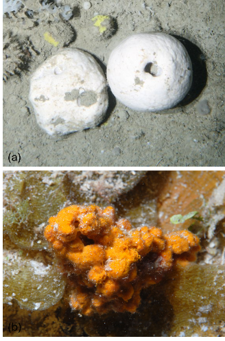
Figure 1. (a) Geodia barretti (b) Axinella corrugata
The cells of the two distinct species were stained with different fluorescent dyes and subsequently mixed. Polyethylene glycol was employed to merge the cells, and Fluorescence-Activated Cell Sorting ( (FACS) was used to isolate and create a homogeneous population of fused cells.
The results indicated a staining efficiency of nearly 90% and a fusion efficiency of approximately 5%. After fusion and selection, the cells were cultured and successfully began to divide. These important findings give hope that hybridoma technology can one day be established with higher fusion efficiency rates, producing hybrid sponge cells that not only produce the compound of interest but also divide.
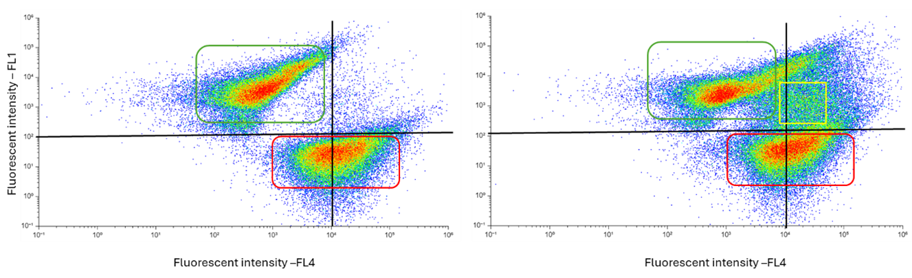
Figure 2. Dot plots obtained from flow cytometry. The left image shows a sample with mixed cells of G. barretti and A. corrugata, while the right image shows the fused cells. The x-axis (FL1) represents the fluorescence intensity of one stain, and the y-axis (FL4) represents the fluorescence intensity of the other stain. In the mixed sample, two populations appear, one for G. barretti cells and one for A. corrugata cells. In the fused sample, a third population appears between the two, indicating cells with both fluorescence signals. Both axes are presented on a logarithmic scale.